Drop Dead!

You need your character to unexpectedly drop dead, with no warning. You need a good, plausible medical reason for this to happen.

You’ve come to the right place, evil killer of beloved characters! ::ahemGeorgeRRMartincough::

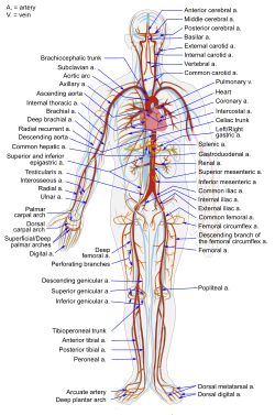
Note: It might help to review “I ❤ the Lub-Dubber”, the post about the cardiovascular system.

Another note: While you’re reading this, remember that the first priority of a first responder to a medical emergency is their ABCs: Airway, Breathing, and Circulation. This is because the body cannot survive without proper oxygenation. Now, without further ado:

Here are six medical conditions that could cause your character to suddenly drop dead.

Aortic Dissection:

What it is: The aorta is the main artery that leads out of the heart. Like all arteries, its walls are made up of multiple layers. Sometimes, a tear occurs in the inner layer. This allows the high-pressure blood in the aorta to push itself in between the layers, “dissecting” the inner layer away from the other layers.

Why the character will die: There are a couple of problems that this can cause. A main one: the flap of the inner layer can cover up the openings to arteries that lead out of the aorta. This can lead to ischemia (oxygen deprivation) of whatever was at the other end of the artery, including the brain or even the heart itself.

Make it plausible: Your character probably has high blood pressure. They might describe a sudden onset of incredibly severe, “tearing” chest pain, which classically radiates to the back.

Hypertrophic Cardiomyopathy:

What it is: A genetic disorder of the heart muscle causes the walls of the left ventricle to get really big and thick. (hyper = over/above, troph = grow, card = heart, myo = muscle, path = feeling/suffering).

Why the character will die: There are a number of ways this can cause problems. (1) In one set of problems, the passage to the aorta (also known as the left ventricular outflow tract) gets blocked by the oversized walls of the left ventricle. No blood gets out of the heart, and therefore nothing in the body can get oxygen. This may happen when they’re at rest, but more often happens when they’re exercising. (2) The electrical system of the heart may be out of whack; that would make it prone to arrhythmias. (See the section on Cardiac Arrhythmia) (3) The heart muscle itself could be less-than-functional: either it doesn’t pump well, or it’s bad at relaxing so it can’t fill up after it pumps.

Make it plausible: Your character is a young, fit athlete. He or she suddenly drops dead while exercising. A member of their family (a parent, for example) may have died suddenly due to the same reason.

Massive Pulmonary Embolism:

What it is: Something blocks off the circulation of blood into the lungs. (Pulmonary = lungs; Embolism = the blockage of a blood vessel with some kind of material.) It’s often a blood clot, but it can also be fat, air, or even tumor material.

Why the character will die: The blood will get backed up from the blockage. This causes a severe drop in blood delivery (measured by blood pressure) to the body, with an increase of pressure behind the blockage. This can cause failure of the right heart (which can’t pump against that kind of pressure). No blood oxygenation, and compromised circulation. Bad news.

Make it plausible:
Emboli that come from blood clots are often from deep vein thromboses, or clots that form in the deep veins (often of the legs). There are some risk factors which make it more likely that your character may develop a pulmonary embolus. These include recent immobilization, recent surgery, stroke, paralysis, chronic heart disease, and cancer. Your character may be obese, may have high blood pressure, and may be a smoker.

Cardiac Arrhythmia:

What it is: A disturbance in the electrical pathways in the heart. This can cause the interruption of proper coordinated pumping in the heart muscle; some arrhythmias can lead to sudden death.

Why the character will die: The pumping of the heart muscle becomes uncoordinated. This results in inadequate filling and/or emptying of the chambers of the heart. Which means that the heart can’t properly pump blood out to the rest of the body. Which means the rest of the body doesn’t get any oxygen.

Make it plausible: There are a LOT of reasons why people get arrhythmias. Everything from being born with extra pathways that run the electrical activity of the heart in a circle, to the sequelae (after-effects) of a heart attack, to drug toxicity, to irregularities of ion flow in the cells of the heart. Research is your friend in this case.

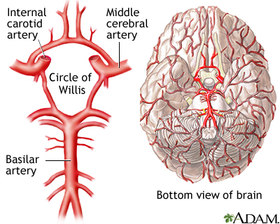
Berry Aneurysm, Sub-Arachnoid Hemorrhage:

What it is: One of the blood vessels in the brain, most likely in the Circle of Willis…

…develops an out-pouching called an aneurysm.

Your character may have been born with it, or they may have acquired it at some point during their life. The acquired ones seem to be more likely to cause problems.

Why the character will die: The aneurysm itself probably doesn’t make much trouble…but it runs the risk of bursting, causing a major brain bleed. The resulting bleed is called a sub-arachnoid hemorrhage, after the space that your character bleeds into. Note: I hyphenated “sub-arachnoid” to make it easier to read and understand. (sub = under, and “arachnoid” refers to the arachnoid mater that surrounds the brain tissue.) It’s usually written as “subarachnoid”. Sub-arachnoid hemorrhages are dangerous; around 50% are fatal.

See that white starfish shape in the middle of the brain? That ain't supposed to be there. That's blood in the arachnoid space. Seeing that on a head film would sure squeeze the heck out of your adrenals (as my cardiology preceptor would say).

Make it plausible: Your character will likely have high blood pressure, and will probably smoke cigarettes. Your character may also be a heavy drinker. A family member (parent, sibling, grandparent) may also have died suddenly from the same cause, but not always.

Are there any that AREN’T cardiovascular?

Cardiovascular causes do seem to be pretty prominent, don’t they? Strokes, aneurysms, emboli, heart attacks…they all have to do with the heart and blood vessels.

The only non-cardiovascular cause of potentially instant-ish death I can think of off the top of my head is anaphylaxis. Which, um, has a cardiovascular cause of death. ::sigh:: I’ll include it anyway.

Anaphylaxis

What it is: Anaphylaxis is an overwhelming allergic reaction to a triggering factor. The substance can be anything from a bee-sting to a food item to a medication. (I couldn’t find any pictures that satisfied me for this condition; there’s just too much going on to accurately represent it with one image.)

Why the character will die: There are two main problems that may result in death from anaphylaxis. (1) There is a certain balance of where the fluid in your body likes to stay, be it in blood vessels or in the general body tissues. In anaphylaxis, there is a massive shift of fluid out of the blood vessels and into the body tissues. That results in the compromise of the cardiovascular system (can’t escape it, can we?), which endangers the perfusion of oxygen to the brain and tissues. (2) There is a significant amount of tissue swelling involved, and this can extend to the breathing passages. This results in a blockage of the airway. That means that the character will not be able to get enough air into their lungs to properly oxygenate their blood. And you have the same problem.

Make it plausible: A food allergy will often result in airway compromise and respiratory arrest. A venom allergy (beesting, etc.) will often result in cardiovascular compromise. Also, there are some age correlations with triggering factors, though pretty much anyone can react to pretty much anything regardless of age. That said,
–Adolescents and young adults are at most risk of death from food reactions.
–Middle-aged adults are at most risk of death from venom reactions
–Older adults are at most risk of death from drug reactions


Last note today, I promise: Any one of these can be fatal or not, depending on the person and the situation.
Sell it how you want to sell it, but make sure you set up the situation properly for plausibility purposes.

Pictures:

References:

Singer, et al. Unruptured Intracranial Aneurysms. UpToDate Online. September 2009. http://www.uptodate.com/online/content/topic.do?topicKey=cva_dise/8216&selectedTitle=1~13&source=search_result

van Gijn, Jan, et al. Subarachnoid Hemorrhage. Lancet; 1/27/2007, Vol. 369 Issue 9558, p306-318

Manning, Warren J. Clinical Manifestations and Diagnosis of Aortic Dissection. September 2009. http://www.uptodate.com/online/content/topic.do?topicKey=vascular/14665&selectedTitle=1~110&source=search_result

Thompson, et al. Overview of Acute Pulmonary Embolism. UpToDate Online. September 2009. http://www.uptodate.com/online/content/topic.do?topicKey=ven_pulm/12962&selectedTitle=1~150&source=search_result

Levy, et al. Arrhythmia Management for the Primary Care Clinician. UpToDate Online. September 2009. http://www.uptodate.com/online/content/topic.do?topicKey=carrhyth/29474&selectedTitle=3~150&source=search_result

Elliot, et al. Diagnosis and Evaluation of Hypertrophic Cardiomyopathy. UpToDate Online. September 2009. http://www.uptodate.com/online/content/topic.do?topicKey=myoperic/7327&selectedTitle=2~150&source=search_result

Bock, S. Allen. Fatal Anaphylaxis. UpToDate Online. September 2009. http://www.uptodate.com/online/content/topic.do?topicKey=anaphyla/5402&selectedTitle=5~150&source=search_result

The contents of this site, such as text, graphics, images, and other material contained on the Site (“Content”) are for informational purposes only. The Content is not intended to be a substitute for professional medical advice, diagnosis, or treatment. Always seek the advice of your physician or other qualified health provider with any questions you may have regarding a medical condition. Never disregard professional medical advice or delay in seeking it because of something you have read on this Site!

If you think you may have a medical emergency, call your doctor or 911 immediately. This blog does not recommend or endorse any specific tests, physicians, products, procedures, opinions, or other information that may be mentioned on the Site. Reliance on any information provided by this blog, or other visitors to the Site is solely at your own risk.

The Site may contain health- or medical-related materials that are sexually explicit. If you find these materials offensive, you may not want to use our Site. The Site and the Content are provided on an “as is” basis.

If you use this as if it were real medical information, these posts will be published by dead drop. You probably don’t have access to the drop point.

Tools for the Toolbox: I <3 The Lub-Dubber!

“(The heart), nurtured in the seas of rebounding blood,
where most especially is what is called thought by humans,
for the blood round the heart in humans is thought.”
–Empedocles

If you held a gun to my head and asked me what my favorite organ was, I’d probably crap my pants and beg you not to shoot me.

But if you were really serious about getting an answer to your question, I’d probably tell you that I absolutely love the heart. You know, after you took the gun away and I got cleaned up a little bit.

Not just because it pulled a fast one on some of my favorite philosophers. Though I gotta admit, I do love watching the history of knowledge unfold through trial-and-error. But seriously, I think it’s functionally and aesthetically the prettiest organ in the body.

(Kidneys are cool, too, but I get a complex about brainless objects that are nonetheless smarter than I am.)

Wanna see something beautiful?

This shows an MRI of a heart beating. Just watch it for a minute. It’s gorgeous. PS: Can you identify the piano piece? Thanks, Interwebz!

Your heart is basically a chunk of muscle that contracts in a really cool way to push your blood around your body. The heart is the center of the circulatory system. That’s the system that’s responsible for getting important groceries from one place to the other in your body: glucose (cell food), oxygen, etc. It also takes out the trash: urea, carbon dioxide, and all those other yuckies that would otherwise gum up the works. And poison you and stuff. Basically, anything that needs to get from one place to another goes through the circulatory system.

It’s like the internet and the transportation system combined: it’s a series of tubes! And through the tubes goes everything from gasses to sugars to hormonal messages.

What’s the setup for the body’s circulation?

The circulatory system is organized like an infinity sign or a figure-eight, with the heart at the cross-over point. One loop is the pulmonary loop that goes to the lungs, and the other is the systemic loop that goes to the rest of the body and all of the organs.

Okay, now we’re red blood cells! We’re going to take a trip through the circulatory system!

Can we go to Hawaii instead?

No.

Why not?

Because we’re red blood cells. And we’re going to take a trip through the circulatory system.

There are red blood cells in Hawaii. And, if I recall correctly, there are also circulatory systems in Hawaii.

. . .

. . . You’re right.

Yes. Yes, I am.

Okay, then. We’re red blood cells. In Hawaii.

Hooray!

Let’s arbitrarily start our journey right before we get into the heart.

Our job as red blood cells is to carry oxygen (O2). We’ve just popped the O2 off of our hemoglobin at our last stop, and loaded up with carbon dioxide (CO2). We want to get the CO2 out of the body, and pick up some more oxygen so we can make another delivery.

We enter the heart through a big vein called the vena cava. The vena cava empties into the first chamber of the heart, the right atrium.

The right atrium is a little room made of contracting muscle. Its job is to top off the right ventricle, which is the main pumping chamber of the right side of the heart. The atrium and ventricle are separated by a one-way “door” called the tricuspid valve (so called because it has three “cusps”, or leaflets.)

So, the right atrium is going to give a squeeze, and propel us through the tricuspid valve into the right ventricle. The right ventricle is relaxing after its previous squeeze; that’s called “diastole”.

It’s very. . . um. . . muscular in here.

Yeah, these pumping chambers are made completely of bundles of muscle fibers. That’s important, because the heart needs to put some force behind its squeezes.

The right ventricle’s job is to provide the force that pushes us through to the lungs, so we can exchange our load of CO2 for O2. To get there, we have to go through another one-way “door” called the pulmonic valve, and through a series of splitting tubes called the pulmonary arteries.

Note: Some people are taught that “arteries carry oxygenated blood and veins carry de-oxygenated blood.” This isn’t entirely correct. By definition, arteries carry blood away from the heart and veins carry blood toward the heart. In the pulmonary (lung) circulation, arteries carry de-oxygenated blood, and veins carry oxygenated blood.

So the right ventricle gives a good squeeze! This is called “systole”. And now, we shoot through the pulmonic valve. We travel through the pulmonary arteries, and end up in the tree-branch-like system of the capillaries of the lungs.

These capillaries are tiny blood vessels that are only wide enough to let one blood cell through at a time. So let’s hold hands so we don’t lose each other, and squeeze on through. We’re close enough to the alveoli (the tiny bags that fill with air when you breathe) that we can drop off our CO2, and load up some O2 for our next delivery run.

Alveoli

And now we’re set to move on!

After we traverse the capillaries, we find ourselves in a reverse-tree-branch-like system: the tiny capillaries are coalescing to form larger and fewer vessels, called the pulmonary veins. We’re on our way back to the heart!

When we reach the “trunk of the tree”, we’re in the pulmonary vein. This empties into the left atrium. The function of the left atrium is a lot like the function of the right atrium. Its job is to top off the left ventricle.

The one-way “door” between the left atrium and the left ventricle is called the mitral valve. So, the left atrium is going to give a good squeeze, and push us through the mitral valve into the left ventricle. The left ventricle is relaxing; it’s in diastole.

And we’re stunned. We’re just flabbergasted.

Um…why?

Remember the right ventricle? The main pumping chamber that pushed us into the lungs?

Yeah?

It was pretty strong, right?

Yeah, it gave us a good push. It took some force to do that.

Well, strength-wise, the left ventricle could EAT THE RIGHT VENTRICLE FOR BREAKFAST. You know, not even breakfast. Maybe even just a midmorning snack. The left ventricle is HUGE!

Look how much bigger the Left Ventricle is on cross-section!

Maybe it’s compensating for something.

Yeah, it is! It’s compensating for the fact that it has to provide enough force to squeeze blood THROUGH THE ENTIRE BODY!

eep.

So we’ve just come through the mitral valve, and we’re chilling in the left ventricle. In front of us, we can see the aortic valve, which is the one-way “door” that leads out of the heart into the main artery of the body, the aorta.

And I can feel the left ventricle getting ready to squeeze. Get ready for systole!

Um . . .

It’s building up! It’s gonna be a big one!

Ummm. . .

What? What’s wrong?

Stop the circulatory system! I wanna get off!

Too late! Hold on, we’re into SYSTOLEEEEEEEEEEEEE!!!!!!!!!!!!!!!!!!!

AAAAAAAAAAAAAAAAAAA!!!!!!!!!!!!!!!

Observe my sloppy Photoshopping on the way!

We’re sailing through the aortic valve and into the aorta! We have a lot of places we can go to deliver our oxygen now, and our first choice comes up almost immediately: the coronary arteries, which supply oxygen to the heart muscle. These guys are really important; if they get blocked off, you get a myocardial infarction (myo = muscle, card = heart, infarct = oxygen deprivation), also known as a heart attack.

But there’s plenty of blood heading into the coronary arteries; let’s stick with the aorta for a bit.

The Aorta has many branches; each one is an artery going to a different part of the body. The carotid arteries head up to the brain. The renal arteries come off of the aorta as it descends into the abdomen, and supply the kidneys. Just look at all these options!

So, pick a part of the body, and we’ll go there and dump our O2. Then we’ll pick up any CO2 it needs to get rid of.

Let’s go to the fourth toe on the right foot.

Um…sure. Good a place as any, I guess. Any particular reason?

How often does that guy get any attention?

Okay, fair enough. So we’re going to travel through the arterial tree, traveling through smaller and smaller arteries until we reach the bed of capillaries that supplies the (ahem) fourth toe on the right foot. Each cell is within striking distance of a capillary, so it can have access to supplies and waste removal services.

Now pick a cell and unload your O2 so it can keep doing its job. And go ahead and pick up some CO2, too.

Got it. Let’s blow this popsicle stand.

Awesome!

So, now we’re going to head out the other side of the capillary bed. The blood vessels are coalescing again, making bigger and bigger vessels. This is the venous part of the systemic circuit.

venous?

Venous.

Venous!

Hee hee! VENOUS!

VENOUS!

Okay, okay, you win. I can’t shout any louder without getting people confused as to who’s talking.

Ooh, self-referential font humor. I’ll declare victory anyway.

Good job.

Okay, we’re headed through the veins on the way back to the heart. The veins coalesce into the vena cava. . . and we’re back where we started.

From here, we’d pass through the right heart into the pulmonary circuit, and then pass through the left heart into the systemic circuit, and around and around and around.

But that would make me a little dizzy, and plus, this post is a monster as it is.

So, that’s the mechanical setup of the circulatory system. It’s just a series of pumps and pipes. If you want to mess with it. . . just think about what happens when a system of pipes gets backed up. Or if the valves start allowing back-flow, or if they get too stiff to allow much flow. Or if the pump becomes weak and inefficient. Or if the pressure gets too high for the pump to push against.

But before I sign off for today, wanna see something gorgeous?

This is the procedure I spent the vast majority of last month observing. It’s called a cardiac catheterization. We’re taking pictures of the coronary arteries by shooting a little bit of opaque dye into them under an X-ray machine. This test looks for blocked or narrowed coronary arteries, which could cause a heart attack if they’re not opened up.

Man, I could look at those forever.

What’s your favorite organ system?

Source:

Cohen, Curd, and Reeve. Readings in Ancient Greek Philosophy. Second edition. Hackett Publishing Company, 2000.

Pictures:

The contents of this site, such as text, graphics, images, and other material contained on the Site (“Content”) are for informational purposes only. The Content is not intended to be a substitute for professional medical advice, diagnosis, or treatment. Always seek the advice of your physician or other qualified health provider with any questions you may have regarding a medical condition. Never disregard professional medical advice or delay in seeking it because of something you have read on this Site!

If you think you may have a medical emergency, call your doctor or 911 immediately. This blog does not recommend or endorse any specific tests, physicians, products, procedures, opinions, or other information that may be mentioned on the Site. Reliance on any information provided by this blog, or other visitors to the Site is solely at your own risk.

The Site may contain health- or medical-related materials that are sexually explicit. If you find these materials offensive, you may not want to use our Site. The Site and the Content are provided on an “as is” basis.

If you use this as if it were real medical information, I’ll take my circulatory system to Hawaii without you.

Published in: on March 11, 2010 at 6:00 am  Comments (28)  
Tags: , , , ,