“(The heart), nurtured in the seas of rebounding blood,
where most especially is what is called thought by humans,
for the blood round the heart in humans is thought.” –Empedocles
If you held a gun to my head and asked me what my favorite organ was, I’d probably crap my pants and beg you not to shoot me.
But if you were really serious about getting an answer to your question, I’d probably tell you that I absolutely love the heart. You know, after you took the gun away and I got cleaned up a little bit.
Not just because it pulled a fast one on some of my favorite philosophers. Though I gotta admit, I do love watching the history of knowledge unfold through trial-and-error. But seriously, I think it’s functionally and aesthetically the prettiest organ in the body.
(Kidneys are cool, too, but I get a complex about brainless objects that are nonetheless smarter than I am.)
Wanna see something beautiful?
This shows an MRI of a heart beating. Just watch it for a minute. It’s gorgeous. PS: Can you identify the piano piece? Thanks, Interwebz!
Your heart is basically a chunk of muscle that contracts in a really cool way to push your blood around your body. The heart is the center of the circulatory system. That’s the system that’s responsible for getting important groceries from one place to the other in your body: glucose (cell food), oxygen, etc. It also takes out the trash: urea, carbon dioxide, and all those other yuckies that would otherwise gum up the works. And poison you and stuff. Basically, anything that needs to get from one place to another goes through the circulatory system.
It’s like the internet and the transportation system combined: it’s a series of tubes! And through the tubes goes everything from gasses to sugars to hormonal messages.
What’s the setup for the body’s circulation?
The circulatory system is organized like an infinity sign or a figure-eight, with the heart at the cross-over point. One loop is the pulmonary loop that goes to the lungs, and the other is the systemic loop that goes to the rest of the body and all of the organs.

Okay, now we’re red blood cells! We’re going to take a trip through the circulatory system!
Can we go to Hawaii instead?
No.
Why not?
Because we’re red blood cells. And we’re going to take a trip through the circulatory system.
There are red blood cells in Hawaii. And, if I recall correctly, there are also circulatory systems in Hawaii.
. . .
. . . You’re right.
Yes. Yes, I am.
Okay, then. We’re red blood cells. In Hawaii.
Hooray!
Let’s arbitrarily start our journey right before we get into the heart.
Our job as red blood cells is to carry oxygen (O2). We’ve just popped the O2 off of our hemoglobin at our last stop, and loaded up with carbon dioxide (CO2). We want to get the CO2 out of the body, and pick up some more oxygen so we can make another delivery.
We enter the heart through a big vein called the vena cava. The vena cava empties into the first chamber of the heart, the right atrium.

The right atrium is a little room made of contracting muscle. Its job is to top off the right ventricle, which is the main pumping chamber of the right side of the heart. The atrium and ventricle are separated by a one-way “door” called the tricuspid valve (so called because it has three “cusps”, or leaflets.)

So, the right atrium is going to give a squeeze, and propel us through the tricuspid valve into the right ventricle. The right ventricle is relaxing after its previous squeeze; that’s called “diastole”.
It’s very. . . um. . . muscular in here.
Yeah, these pumping chambers are made completely of bundles of muscle fibers. That’s important, because the heart needs to put some force behind its squeezes.
The right ventricle’s job is to provide the force that pushes us through to the lungs, so we can exchange our load of CO2 for O2. To get there, we have to go through another one-way “door” called the pulmonic valve, and through a series of splitting tubes called the pulmonary arteries.
Note: Some people are taught that “arteries carry oxygenated blood and veins carry de-oxygenated blood.” This isn’t entirely correct. By definition, arteries carry blood away from the heart and veins carry blood toward the heart. In the pulmonary (lung) circulation, arteries carry de-oxygenated blood, and veins carry oxygenated blood.
So the right ventricle gives a good squeeze! This is called “systole”. And now, we shoot through the pulmonic valve. We travel through the pulmonary arteries, and end up in the tree-branch-like system of the capillaries of the lungs.

These capillaries are tiny blood vessels that are only wide enough to let one blood cell through at a time. So let’s hold hands so we don’t lose each other, and squeeze on through. We’re close enough to the alveoli (the tiny bags that fill with air when you breathe) that we can drop off our CO2, and load up some O2 for our next delivery run.

Alveoli
And now we’re set to move on!
After we traverse the capillaries, we find ourselves in a reverse-tree-branch-like system: the tiny capillaries are coalescing to form larger and fewer vessels, called the pulmonary veins. We’re on our way back to the heart!

When we reach the “trunk of the tree”, we’re in the pulmonary vein. This empties into the left atrium. The function of the left atrium is a lot like the function of the right atrium. Its job is to top off the left ventricle.

The one-way “door” between the left atrium and the left ventricle is called the mitral valve. So, the left atrium is going to give a good squeeze, and push us through the mitral valve into the left ventricle. The left ventricle is relaxing; it’s in diastole.
And we’re stunned. We’re just flabbergasted.
Um…why?
Remember the right ventricle? The main pumping chamber that pushed us into the lungs?
Yeah?
It was pretty strong, right?
Yeah, it gave us a good push. It took some force to do that.
Well, strength-wise, the left ventricle could EAT THE RIGHT VENTRICLE FOR BREAKFAST. You know, not even breakfast. Maybe even just a midmorning snack. The left ventricle is HUGE!

Look how much bigger the Left Ventricle is on cross-section!
Maybe it’s compensating for something.
Yeah, it is! It’s compensating for the fact that it has to provide enough force to squeeze blood THROUGH THE ENTIRE BODY!

eep.
So we’ve just come through the mitral valve, and we’re chilling in the left ventricle. In front of us, we can see the aortic valve, which is the one-way “door” that leads out of the heart into the main artery of the body, the aorta.
And I can feel the left ventricle getting ready to squeeze. Get ready for systole!
Um . . .
It’s building up! It’s gonna be a big one!
Ummm. . .
What? What’s wrong?
Stop the circulatory system! I wanna get off!
Too late! Hold on, we’re into SYSTOLEEEEEEEEEEEEE!!!!!!!!!!!!!!!!!!!
AAAAAAAAAAAAAAAAAAA!!!!!!!!!!!!!!!

Observe my sloppy Photoshopping on the way!
We’re sailing through the aortic valve and into the aorta! We have a lot of places we can go to deliver our oxygen now, and our first choice comes up almost immediately: the coronary arteries, which supply oxygen to the heart muscle. These guys are really important; if they get blocked off, you get a myocardial infarction (myo = muscle, card = heart, infarct = oxygen deprivation), also known as a heart attack.
But there’s plenty of blood heading into the coronary arteries; let’s stick with the aorta for a bit.

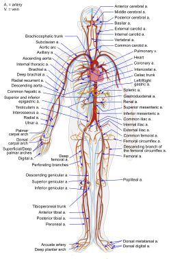
The Aorta has many branches; each one is an artery going to a different part of the body. The carotid arteries head up to the brain. The renal arteries come off of the aorta as it descends into the abdomen, and supply the kidneys. Just look at all these options!

So, pick a part of the body, and we’ll go there and dump our O2. Then we’ll pick up any CO2 it needs to get rid of.
Let’s go to the fourth toe on the right foot.
Um…sure. Good a place as any, I guess. Any particular reason?
How often does that guy get any attention?
Okay, fair enough. So we’re going to travel through the arterial tree, traveling through smaller and smaller arteries until we reach the bed of capillaries that supplies the (ahem) fourth toe on the right foot. Each cell is within striking distance of a capillary, so it can have access to supplies and waste removal services.
Now pick a cell and unload your O2 so it can keep doing its job. And go ahead and pick up some CO2, too.
Got it. Let’s blow this popsicle stand.
Awesome!

So, now we’re going to head out the other side of the capillary bed. The blood vessels are coalescing again, making bigger and bigger vessels. This is the venous part of the systemic circuit.
venous?
Venous.
Venous!
Hee hee! VENOUS!
VENOUS!
Okay, okay, you win. I can’t shout any louder without getting people confused as to who’s talking.
Ooh, self-referential font humor. I’ll declare victory anyway.
Good job.
Okay, we’re headed through the veins on the way back to the heart. The veins coalesce into the vena cava. . . and we’re back where we started.

From here, we’d pass through the right heart into the pulmonary circuit, and then pass through the left heart into the systemic circuit, and around and around and around.
But that would make me a little dizzy, and plus, this post is a monster as it is.
So, that’s the mechanical setup of the circulatory system. It’s just a series of pumps and pipes. If you want to mess with it. . . just think about what happens when a system of pipes gets backed up. Or if the valves start allowing back-flow, or if they get too stiff to allow much flow. Or if the pump becomes weak and inefficient. Or if the pressure gets too high for the pump to push against.
But before I sign off for today, wanna see something gorgeous?
This is the procedure I spent the vast majority of last month observing. It’s called a cardiac catheterization. We’re taking pictures of the coronary arteries by shooting a little bit of opaque dye into them under an X-ray machine. This test looks for blocked or narrowed coronary arteries, which could cause a heart attack if they’re not opened up.
Man, I could look at those forever.
What’s your favorite organ system?
Source:
Cohen, Curd, and Reeve. Readings in Ancient Greek Philosophy. Second edition. Hackett Publishing Company, 2000.
Pictures:
.svg/350px-Diagram_of_the_human_heart_(cropped).svg.png)


The contents of this site, such as text, graphics, images, and other material contained on the Site (“Content”) are for informational purposes only. The Content is not intended to be a substitute for professional medical advice, diagnosis, or treatment. Always seek the advice of your physician or other qualified health provider with any questions you may have regarding a medical condition. Never disregard professional medical advice or delay in seeking it because of something you have read on this Site!
If you think you may have a medical emergency, call your doctor or 911 immediately. This blog does not recommend or endorse any specific tests, physicians, products, procedures, opinions, or other information that may be mentioned on the Site. Reliance on any information provided by this blog, or other visitors to the Site is solely at your own risk.
The Site may contain health- or medical-related materials that are sexually explicit. If you find these materials offensive, you may not want to use our Site. The Site and the Content are provided on an “as is” basis.
If you use this as if it were real medical information, I’ll take my circulatory system to Hawaii without you.

Fascinating, thanks!
So now I’m curious how that opaque dye in the last video works. Is it coating the inside of the arteries, or just flooding them with dye so that they stay visible? Why aren’t the veins also lighting up?
Also, what goes on when, say, the patient is in a high CO2/CO atmosphere? Does anything different happen, or do you just wind up with a lot of CO2-saturated red blood vessels going through the body? (Not that I have a murder mystery plot riding on this or anything…)
So. You enter the arterial system through the femoral artery in the leg, and run a catheter (a small tube) against the flow, up into the aorta to the origins of the coronary arteries. Then you flood brief squirts of dye into the arteries to make them visible.
You don’t see the veins light up because the picture cuts away right after the arteries are adequately seen. After the picture cuts out, the dye filters through the capillary beds, and then into the veins like you said it should.
I wrote a post recently on oxygen transport, which might begin to (incompletely) answer your question. You can find it here: https://doctorgrasshopper.wordpress.com/2010/02/06/superballs-pockets-and-fun-with-awesome-molecules/
I’ll probably get around to writing a post on cellular respiration at some point. It’s a really cool topic, and I’m glad you brought it up! I hope you can be patient with me, though…I’m backed way up as it is!
Thanks for reading!
Ah, how did I miss that? Thanks. I’m more interested in CO2, but that looks like a good starting point for research on the subject.
Awesome post! You make medical stuff interesting! And, you know, that’s a major compliment, even from me, who usually finds medical stuff fascinating. 🙂
One thing you didn’t touch on was what makes the heart muscles move. The plumbing is important, but so is the electrical system! Not that it’s my favourite system, or anything, but it’s very important to some plots I have. And I’m interested in the way it affects the heart, too.
Thanks so much for all this information! Have I mentioned that this blog is exactly what I’ve been looking for in my writing research? Thank you!
So glad you like the blog, BJ! Thanks for your encouragement!
Yeah, I know I didn’t touch heart electrophysiology in this post. I kinda did that on purpose. It’s a really, really neat topic, and I think it deserves its own post…eventually. It’s pretty complex, though, and I’m going to need some time to think about how to present it so no one ends up bashing their heads against the wall (including me! 😀 )
If you want to get a jump on the topic, there’s a relatively straightforward book called Rapid Interpretation of EKGs, by Dale Dubin. (I have the 6th edition, published in 2000 by Cover Inc.) I didn’t read it until after I had taken physiology, but I think it should be pretty accessible to educated laypeople.
Hope that can tide you over until I get around to talking about it over here!
Missed this response. Sorry. I may have to look for that book… or just wait for your post. 🙂 It’s not something I’m facing right now in my writing, so waiting may be worthwhile.
Thanks!
Paul Schwartz – Cafe del Mar – Aria 2 – 06 - Cantilena
Thanks, atsiko! You rock!
http://www.paulschwartz.com/aria2_c_cantilena.html
What’s your favorite organ system?
How about the eye? That’s a pretty cool organ. Or maybe the vestibular system in the ears? That’s awesome too.
Indeed!
Brain all the way. We’re writers, aren’t we?
Most of the writers I’ve known are more concerned about their stomachs 😉
The more fool they. I live on stomach acid.
Brains are definitely cool! And if you’re a zombie, you can be concerned about both brains and your stomach at the same time!
Yes, but you also have to worry about shotguns and people with metal plates in their heads. Poor zombie teeth.
You talked about the difference between veins and arteries (carrying oxygenated blood, etc.) I was also told that veins use the body’s muscle contractions to move blood, while arteries rely on that really cool left ventricle. Do the pulmonary veins work the same way?
That’s a really good question, C. To be honest, I don’t rightly know off the top of my head. My guess is that the movement of the lungs expanding and contracting helps move the blood along in the pulmonary circulation.
I know there are a couple of real doctors who have been stopping by……anyone know the answer?
[…] It might help to review “I ❤ the Lub-Dubber”, the post about the cardiovascular […]
did u know you’r awesome, because you are. Thanks alot this really helped. I really enjoyed the humorous comments as i read along, thanks alot.
You’re too kind! I’m so glad you liked it, and I’m glad I could help you out!
Dr. G
[…] looking at a cross-section of the left ventricle of the heart. The ventricle on the right is relatively normal, and the ventricle on the left is suffering from […]
[…] listen. Gravity is always pulling your blood down toward the center of the earth. Your cardiovascular system is all set up to fight against this force to keep the blood circulating adequately to all parts of […]
I really luv dis.science is the best subject .BAM,DIS IS CRAZY.GOD bless u man and GOD bless science and technology
Well thanks, omojola! I’m glad you stopped by!
aaaaaaaaaaaaaaaaaaaaaaaaaaaaaaaaaaaaaaaaaaaaaaaaaaaaaaaaaaaaaaaaaaaaaaaaaaaaaaaaaaaaaaaaaaaaaaaaaaaaaaaaaaaaaaaaaaaaaaaaaaaaaaaaaaaaaaaaaaaaaaaaaaaaaaaaaaaaaaaaaaaaaaaaaaaaaaaaaaaaaaaaaaaaaaaaaaaaaaaaaaa!!!!!!!!!!!!!!!!!!!!!!!!!!!!!!!!!!!!!!!!!!!!!!!!!!
-___- Wow ? !
[…] Ah. Blog-flashback humor. […]